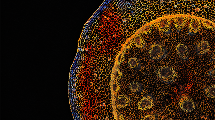
New Spectral Fluorescence Lifetime Imaging Method Developed for Investigating Live Cells
UC Irvine biomedical engineering researchers have developed a new fast, robust microscopy imaging technique that could better capture detailed and precise information of cellular processes, such as characterizing migrating cancer cells. The technique combines two broadly applied microscopy methods – spectral imaging and fluorescence lifetime imaging microscopy (FLIM) – by developing a true parallel detection system for simultaneous measurements that can be processed in real time.
Led by Enrico Gratton, Distinguished Professor of biomedical engineering and director of the Laboratory for Fluorescence Dynamics at the Samueli School of Engineering, the team published their research in the April 15, 2021, issue of Nature Methods.
“This new technique works on live cells; there is no need for fixation,” said Gratton. “We believe the information we obtain could be augmented by genomic and proteomics data. More importantly, Phasor S-FLIM can provide results in tissues that are difficult to measure using these omics approaches. This would be a new paradigm for researchers who are using advanced imaging to solve hard-to-investigate living cells and gain important insights on human health.”
The new fluorescence microscopy method uses the color properties and emission duration of fluorescent dyes to achieve high specificity and sensitivity in imaging living cells. It is an extremely valuable tool in biomedical research as most of today’s microscopes rarely obtain emission spectrum and fluorescence lifetime, and only a handful can do it on the same microscope. This process is, however, very slow and requires high power for illumination, greatly limiting the acquisition speed and damaging the sample.
Read the full article on the UCI Samueli School of Engineering website.


