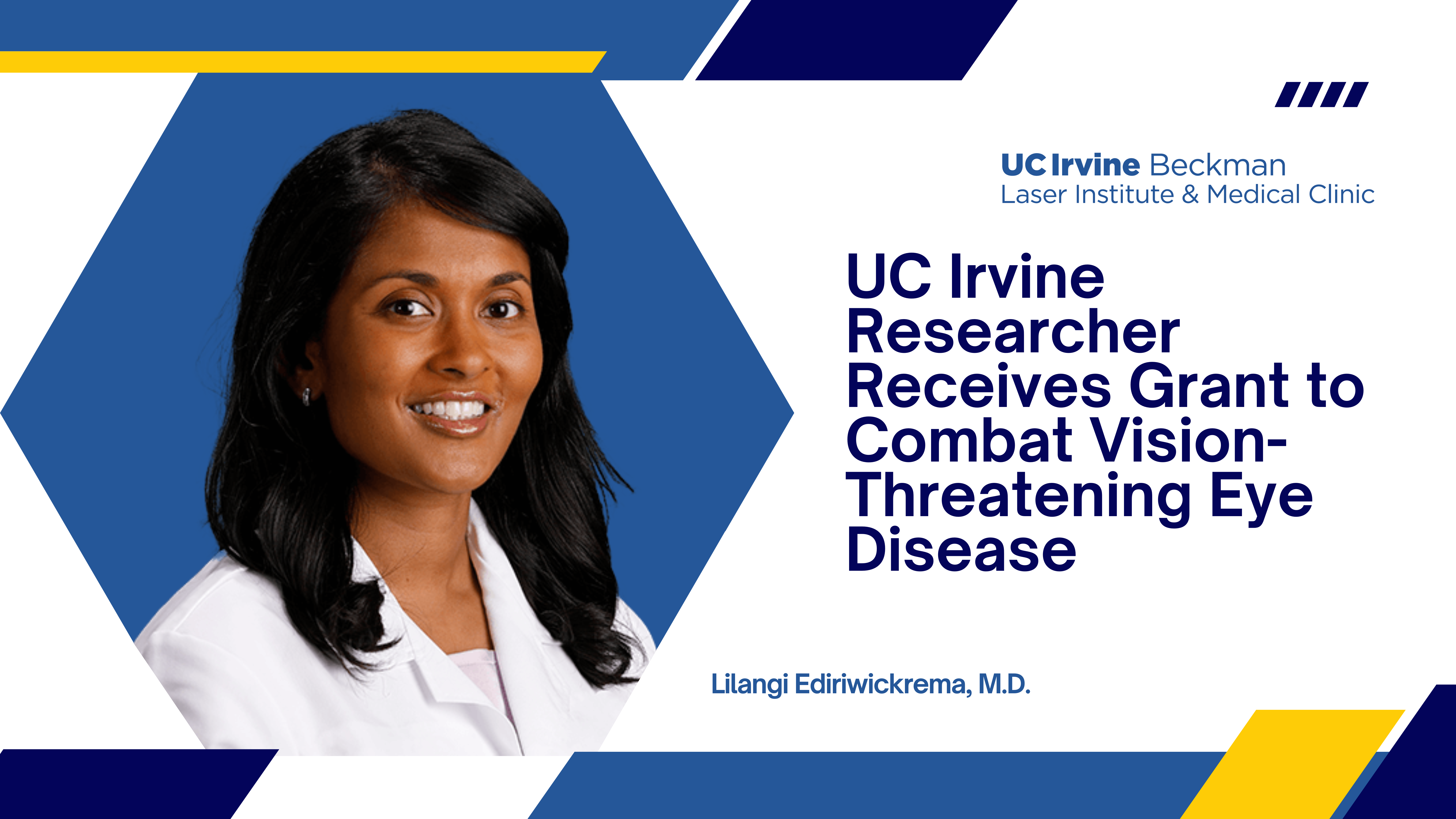
UC Irvine Researcher Receives Grant to Combat Vision-Threatening Eye Disease
Dr. Lilangi Ediriwickrema awarded K23 grant to develop breakthrough imaging technology for thyroid eye disease
Dr. Lilangi Ediriwickrema, a UCI Health ophthalmologist at Gavin Herbert Eye Institute, has been awarded a competitive K23 Mentored Patient‑Oriented Research Career Development Award from the National Eye Institute (NEI) to advance a novel imaging approach for thyroid eye disease (TED). Her project, “Spatial Frequency Domain Imaging to Investigate Mechanistic Constituents in Active Thyroid Eye Disease,” aims to transform how clinicians objectively assess disease activity and treatment response in this complex, vision‑threatening condition.
The project leverages Spatial Frequency Domain Imaging (SFDI), a noninvasive optical imaging technology developed at UC Irvine, to quantify tissue‑level vascular and structural changes associated with TED. The research integrates advanced biomedical optics with patient‑oriented clinical investigation, reflecting a strong collaboration between engineering innovation and ophthalmic care.
A Disease That Goes Beyond Vision
Thyroid eye disease affects an estimated 90 to 300 per 100,000 people globally, with some estimates suggesting up to half a million cases in the United States. Most commonly associated with Graves’ disease, TED occurs when the immune system attacks muscle and fat tissue behind the eyes, leading to painful inflammation, bulging of the eyes, blurred or double vision, and in severe cases, vision loss.
“Moderate to severe thyroid eye disease can also be socially stigmatizing, affecting patients well beyond their clinical symptoms,” explains Dr. Ediriwickrema, assistant professor in the UC Irvine School of Medicine Department of Ophthalmology. “It can profoundly impact quality of life, self‑confidence, and social interactions.”
Pioneering Noninvasive Technology Through Cross‑Disciplinary Mentorship
The study is conducted under the primary mentorship of Dr. Anthony J. Durkin, Director of the SFDI laboratory at UC Irvine and a co‑inventor of the SFDI technique. Dr. Durkin has played a key role in the development and translational application of SFDI for biomedical and clinical imaging, enabling objective, quantitative assessment of tissue physiology.
Technical development, system optimization, and imaging troubleshooting for the project are supported by Dr. Gordon Kennedy, a project scientist and senior member of the SFDI laboratory, whose expertise in optical instrumentation and quantitative imaging methods has been central to adapting SFDI for orbital and peri‑ocular applications relevant to thyroid eye disease.
“Translating optical imaging technologies into meaningful clinical tools requires close collaboration between clinicians and engineers,” said Dr. Durkin. “This project exemplifies how sustained mentorship and advanced imaging infrastructure can support patient‑centered innovation.”
Comprehensive Validation and Clinical Impact
In addition to in‑vivo imaging of patients with active TED and age‑matched healthy controls, the research team will analyze relevant surgical tissue specimens to validate imaging‑derived metrics at a biological level. This combined approach strengthens the potential for SFDI‑based measurements to serve as objective biomarkers of disease activity, severity, and response to therapy. The long‑term goal is to provide clinicians with quantitative tools that can improve diagnosis, guide treatment decisions, and support emerging targeted therapies for thyroid eye disease.
“The treatment landscape is evolving rapidly,” said Dr. Ediriwickrema. “In the coming years, objective imaging biomarkers could play an important role in personalizing care for patients with this disease.”
Training the Next Generation of Clinician‑Scientists
The NEI K23 award supports Dr. Ediriwickrema’s development as an independent clinician‑scientist through a structured mentorship team that includes Drs. Durkin, Bernard Choi, and Anand Ganesan. The award emphasizes patient‑oriented research that directly integrates studies in human subjects with advanced quantitative and imaging methodologies.
“This work reflects UC Irvine’s strength in team‑based translational science,” said Durkin. “By combining clinical insight with innovative optical imaging approaches, we can address important unmet needs in patient care.”
About the National Eye Institute (NEI)
The National Eye Institute (NEI) leads the federal government’s efforts to eliminate vision loss and improve quality of life through vision research, including driving innovation, fostering collaboration, expanding the vision workforce, and educating the public and key stakeholders. The NEI supports basic and clinical science programs to develop sight-saving treatments and to broaden opportunities for people with vision impairment. For more information, visit https://www.nei.nih.gov.
About the National Institutes of Health (NIH)
The National Institutes of Health (NIH), the nation’s medical research agency, includes 27 Institutes and Centers and is a component of the U.S. Department of Health and Human Services. NIH is the primary federal agency conducting and supporting basic, clinical, and translational medical research, and is investigating the causes, treatments, and cures for both common and rare diseases. For more information about NIH and its programs, visit www.nih.gov.

Author: Gabrielle Comfort