Chen, Wong Win NIH Grant to Characterize Mucosal Health

By Anna Lynn Spitzer

UC Irvine biomedical engineering professor Zhongping Chen and otolaryngologist/facial surgeon Dr. Brian Wong recently won a $2.3 million, four-year R01 award from the NIH’s National Institute of Biomedical Imaging and Bioengineering to develop a technology that could help doctors treat sinus and nasal ailments with more precise information. The two, who have collaborated for nearly 20 years, are creating an innovative in vivo imaging system they call a phase-resolved spectrally encoded endoscope (PR-SEE).

Approximately 50 million Americans each year suffer from painful sinus and allergic nasal upper respiratory ailments. Known medically as chronic rhinosinusitis and allergic rhinitis, the resulting headaches, stuffy/runny noses, itchy, watery eyes and sneezing are responsible for health care expenditures of more than $35 billion a year, along with 3.5 million missed work days. “It’s a quality of life issue,” says Wong, who has a joint appointment in the Samueli School’s biomedical engineering department. “You’re probably not going to die from it; you’re just going to be miserable.”

Doctors are limited in their ability to treat these patients, relying mostly on subjective information gleaned from the patients themselves. “Right now, the response to therapy is entirely based on patient-reported outcomes,” says Wong, who adds that it’s difficult to develop appropriate therapies with these “semi-quantitative responses.”

The PR-SEE will employ two distinct imaging techniques that will overcome current limitations on in vivo cilia imaging. (Cilia are the tiny, hairlike structures on airway cell surfaces that sweep in a rhythmic pattern to transport mucus.) The first, optical coherence tomography (OCT), uses two scanning mirrors to provide information from deep within tissue; the second is spectrally encoded interferometry, a method that uses one mirror, providing faster imaging speed, but which cannot achieve deep measurements like OCT. Used together, the two techniques can give medical professionals quantitative information they have not had access to. “This has never been done in vivo before,” Chen says.

The device will measure ciliary beat frequency (CBF) – the speed at which the cilia sweep. Along with other factors, the CBF determines the efficiency of mucus transport, providing a strong indicator of upper airway health.

The device also will assess amplitude and propagation of the mucosal metachronal waves. “CBF is only one of the many factors that dictate the ability of cilia to transport mucus,” explains Chen. “It is also important to study the sweeping pattern (the amplitude) and how well each cilium coordinates with each other (the metachronal waves).

“These quantitative factors – speed/frequency, amplitude, metachronal waves – will provide a more comprehensive understanding of how airway cilia work, and will go a long way toward determining upper airway health,” he says.

In order to obtain functional parameters, Chen and Wong will test their device first on a rabbit nasal airway model, then on anaesthetized patients undergoing nasal surgeries. Eventually, they plan to develop stabilization techniques to help keep non-anaesthetized patients from moving in order to translate the research into a device that can be used in a doctor’s office.

Rhinosinusitis alone currently accounts for more antibiotic prescriptions than any other diagnosis in ambulatory settings, and in more severe cases, results in 600,000-plus sinonasal operations annually.

“Last time I checked, there were over 400 medications approved by the FDA for use in treating the nose. This additional information will help in identifying proper therapies and in determining whether those therapies are working,” Wong says. “We need a rigorous means to measure the response to pharmacological therapy and/or surgery by probing mucosal physiology at a fundamental level.”

Adds Chen: “This imaging modality establishes an objective means to gauge sinus health and the response to treatment, which in turn will aid scientists, clinicians and industry professional to better develop drugs, devices and other therapies.”

Read full article on UCI Samueli School of Engineering website.

Chris Barty is Laser Focused on Laser World Domination


By Jackie Connor, UCI Beall Applied Innovation Main Graphic: Julie Kennedy, UCI Beall Applied Innovation Headline Design: Elisa Le, UCI Beall Applied Innovation UCI’s Distinguished Professor aims to advance the age-old X-ray machine and has uncovered a laser technology that could potentially take the teeth out of COVID-19’s grip. When Chris Barty focuses his mind on something, it is with laser precision. The UC Irvine Distinguished Professor of Physics and Astronomy learned from an early age he was drawn to lasers after watching the first episode of the animated sci-fi adventure series “Jonny Quest.” “Episode one of ‘Jonny Quest’ is the laser episode,” said Barty. “In the episode, [the villains] are burning ships in the Sargasso Sea with a laser… and the episode starts explaining what a laser is.” Post-“Jonny Quest,” Barty received his bachelor’s degree in chemistry, physics and chemical engineering at North Carolina State University and later received his master’s degree and a doctorate in applied physics at Stanford University. Though Barty calls out “Jonny Quest” for his original and perhaps subconscious interest in lasers, Barty’s adventures in the wide world of laser technology are anything but science fiction.

Close-up portrait of the researcher

Graphic: Julie Kennedy, UCI Beall Applied Innovation

Lasers, Lasers, Nothing but Lasers After teaching at Stanford for four years, Barty moved to San Diego to run a privately funded research organization where he focused on intense lasers and X-rays created by intense lasers. In 2000, Barty left San Diego to work as the chief scientific officer at Lawrence Livermore National Lab (LLNL)’s laser division. “I was like a kid in a candy store,” said Barty. “I was doing laser weapons work. I was doing lasers for fusion energy, for clean energy, lasers for medicine, and lasers for nuclear materials detection. The joke about Livermore is that Lawrence Livermore National Lab isn’t what LLNL stands for … it stands for ‘lasers, lasers, nothing but lasers.’” Barty’s division primarily focused on national security, including the use of a laser-Compton system – a laser-based X-ray machine – to recognize Uranium 235 from Uranium 238, two very similar nuclear materials with a major difference. “Uranium 238 is considered ballast but 235 is Hiroshima,” said Barty. “The big worry is that somebody could ship 235 into a port and detonate it and every port in the world could shut down causing a trillion dollar impact on the word economy overnight.” This mission provided his team at LLNL about $70 million in funding to develop the detection technology in addition to another $150 million at the Stanford Linear Accelerator Center (SLAC) to develop an accelerator technology. Thirteen of the patents resulting from the LLNL activities were licensed by Lumitron Technologies and are the foundation of the company.

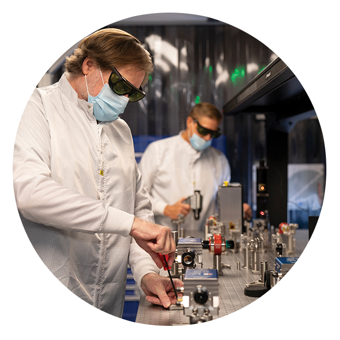
Professor Chris Barty and colleague working in the Lumitron Laser Lab.

Chris Barty, Ph.D., UCI distinguished professor of Physics and Astronomy, work with lasers in Lumitron Technologies’ Laser Lab located in University Research Park in Irvine. Photo: Julie Kennedy, UCI Beall Applied Innovation

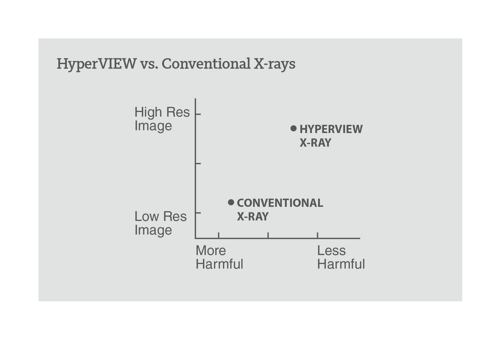
Lumitron Technologies In 2016, Lumitron Technologies was formed as a company that develops and commercializes unique X-ray systems. The company is based on $220 million worth of R&D from LLNL and SLAC. With co-founder and Executive Chairman Maurie Stang, Barty, co-founder, executive director and chief technology officer, decided to set up shop in Irvine. In January, Lumitron completed a $34 million funding round, with the help of Roth Capital Partners in Newport Beach, in addition to $11.6 million from Defense Advanced Research Projects Agency. With the funding, Lumitron aims to build its first commercial X-ray systems. The company is developing a HyperVIEW platform that will provide high-resolution X-ray images, which improves image resolution up to 1,000 times compared to conventional X-rays while, at the same time, imparts a significantly less harmful dose of radiation to the patient – all from a device the size of a modern CT machine. “The impact of Lumitron’s breakthrough will touch a wide array of human endeavors, both in imaging of unsurpassed resolution and new therapies, which will leverage our ability to image and treat simultaneously at a near-cellular level,” said Stang.

Graph showing HyperVIEW provides higher resolution and is less harmful than conventional X-rays

Chart: Rachel Noble, UCI Beall Applied Innovation

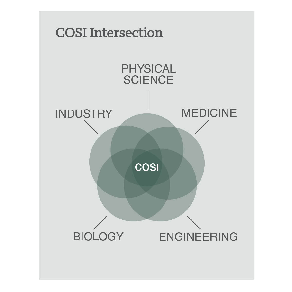
As Barty describes, since the platform’s technology can see down to a cellular level, the potential to detect and treat diseases like cancer can be done in ways that no one has been able to do before. The HyperVIEW platform can detect and treat cancer without introducing radioactive materials to the body. Although commonly used in the medical and research sectors, the technology is also applicable for 3D printing, mining, security, semiconductor manufacturing and nondestructive material evaluation. COSI Lab In the summer of 2017, Barty became the senior faculty member and first hire of UCI’s Convergence Optical Science Initiative (COSI). COSI lab space is located at the Cove @ UCI, UCI Beall Applied Innovation’s headquarters, and is dedicated to studying laser activity – a space he describes as “the intersection of physical science, engineering, biology, medicine and industry all around something photonic.”

Venn diagram showing COSI at the intersection of Physical Science, Medicine, Engineering, Biology, and Industry.

Chart: Rachel Noble, UCI Beall Applied Innovation

“I have a lot of crazy ideas, all the time. That’s what I do,” said Barty. “The academic side is about things that might be two or three or four generations down the road, for how you might either make the machine better or use the machine in a different way.” With Barty’s appointments in the School of Physical SciencesSchool of Medicine and the Beckman Laser Institute and Medical Clinic, he recruits doctorate students to the lab to study the applications of his technologies. Most of what happens in the COSI Lab relates to laser activity, like investigating new applications of common laser technologies. Lasers and COVID-19 Most recently, Barty and his research team at UCI are developing a new technique in the fight against COVID-19 using diode lasers found in a Blu-ray players. The technology can be used as a way to rapidly sterilize surfaces and/or clean the air.

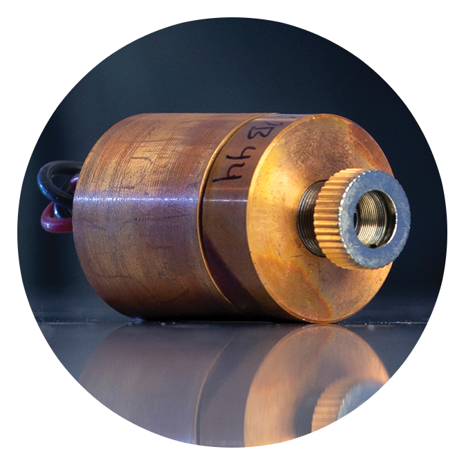
Close-up of a brown diode laser component on a reflective surface.

Pictured is a diode laser found in a Blu-ray player. These small but mighty pieces can be used to sterilize surfaces, clean the air and possibly denature COVID-19. Photo: Steve Zylius

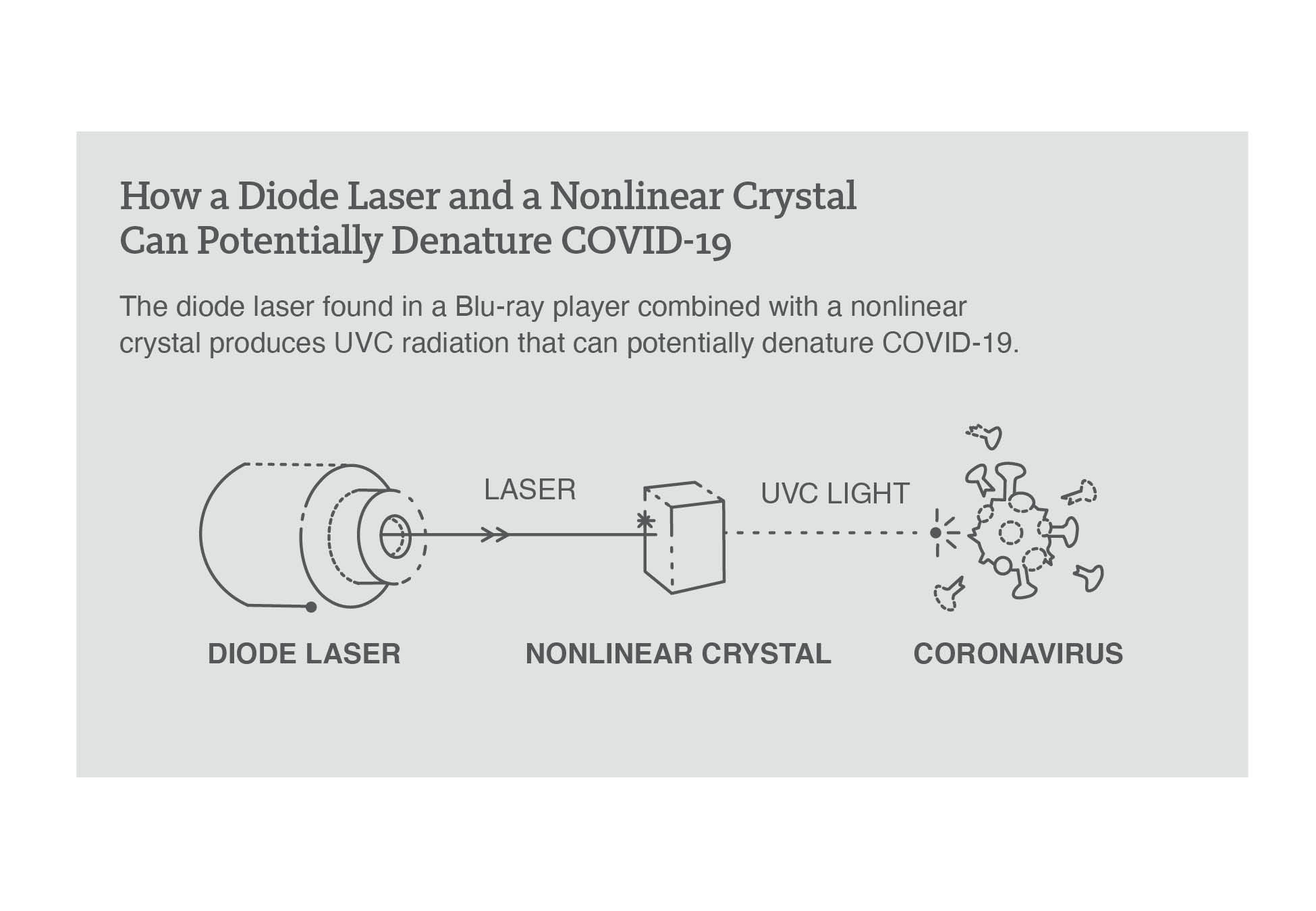
It would be less expensive and safer than mercury discharge lamps used in hospitals, according to Barty, since the mercury lamps emit UVA and UVB light. The technology, which is created by using the laser in the Blu-ray player, produces a shorter wavelength – UVC radiation that can potentially denature COVID-19. “There is medical literature that suggests that short-wavelength UVC is not that bad for you because the dead skin cells that are on your body are enough to absorb the UVC. The teardrop on your eye is enough to absorb very short wavelength UVC,” said Barty. “It doesn’t really damage your skin, your living skin or living tissue in the same way that UVA and UVB does. In practice, it’s not widely used, because it’s expensive.” To create UVC light, it takes $100,000 worth of equipment, according to Barty. However, once Barty and his team put a nonlinear crystal in front of the Blu-ray player’s diode laser, it can create UVC light. The team only needs to spend $50 for the Blu-ray laser diode part. “The semiconductor industry that made the Blu-ray diode lasers has perfected the art of doing that and it’s now become cheap,” said Barty. “They’ve built the fab lines and everything. You’re winning, because somebody else paid the money to do it.”

Diagram showing a diode laser and nonlinear crystal producing UVC light to denature a coronavirus particle.

Graphic: Rachel Noble, UCI Beall Applied Innovation

Barty and his team are currently setting up to investigate the modified laser’s effects on COVID-19. If effective against COVID-19, this technology could be utilized within a building’s air conditioning duct or utilized within light fixtures to constantly clean circulating air. “You could imagine having something where a mask actually has one of these in it,” said Barty. “Every time you take a breath, you’re getting air that has been cleaned, or every time you exhale, all that stuff that you exhale is being cleaned.” Laser-Focused Future Between Lumitron and creating new technologies from the COSI Lab, Barty has one thing in mind for the future of his company: World domination … with lasers, of course. Barty is currently focused on bringing down the cost of the technology so hospitals can easily become equipped with Lumitron’s advanced machines. “I want a Lumitron machine in every hospital,” said Barty. “I want 10 machines in every hospital. My mother died of breast cancer. I’ve got plenty of people in the company who’ve had people that have been impacted by cancer. If we can do anything to solve that, I’m all for it.” Learn more about Lumitron. Read full article in UCI Beall Applied Innovation “Rising Tide.”


Bernard Choi receives seed funding award to plan new center on healthy aging

Bernard Choi, Professor of Biomedical Engineering and Surgery; Susan Charles, Chair and Professor of Psychological Science and Mike Yassa Director of the Center for Neurobiology of Learning and Memory, were awarded seed funding through the Office of Research’s, “Investing to Develop Center-Scale Multidisciplinary Convergence Programs” in support of planning a new center on healthy aging.  The project titled, “Longevity 2.0,” will draw on faculty expertise from multiple UCI Schools to create a dedicated center with research focused on increasing the years of an active, healthy life. The project entails developing partnerships with local community organizations. The seed funding will be used to plan and organize the “Longevity 2.0” center, including creating its infrastructure and identifying core transdisciplinary research aims.

Petra Wilder-Smith receives $973,383 TRDRP award to develop smartphone-based oral scanner pen to detect oral cancer in tobacco users

Dr. Petra Wilder-Smith was awarded a $973,383 Tobacco-Related Disease Research Program (TRDRP) grant to expand their existing artificial intelligence-driven oral cancer probe capabilities to benefit individuals who use tobacco.

To date, Dr. Wilder-Smith and her team have focused on differentiating between cancer and pre-cancer from healthy tissues in the mouth.  However, tobacco users have many intra-oral lesions, which confound the probe in its current configuration.

Their aim is to train the artificial intelligence algorithm to differentiate between the more common mouth lesions, especially tobacco-related ones, as well as cancer, precancer and healthy tissue.  The diagnostic algorithm will be accessed through a simple smartphone or tablet-based App, which also can identify and connect with healthcare providers for communication, remote examination and data transfer.

According to Dr. Wilder-Smith, oral cancer risk in tobacco users is substantially higher compared to non-smokers with low-resource and underserved populations having the highest prevalence of oral cancer and loss of life. At-risk populations commonly lack access to screening, surveillance, specialist access and primary health care with follow-up for the early detection and surveillance that could lead to early treatment and better outcomes in patients.

“More than two-thirds of oral cancers are diagnosed late.  If oral cancer is detected early, then the survival rate in patients vastly increases,” said Dr. Wilder-Smith.  “While the proposed project includes a very simple photographic tool, our overall approach is to overcome the lack of access to specialist diagnosis by high-risk populations through empowering readily available healthcare workers to perform effective oral cancer screenings and surveillance.  We anticipate that this project will directly result in better oral cancer outcomes for high risk populations.”

ABBU(Fatherly) Love!

Dr. Thomas Milner recently appointed as the director of the Beckman Laser Institute at the University of California, Irvine, has been an active member and integral contributor to the Bridge Ventilator Consortium. During the peak of the COVID-19 pandemic, Dr. Milner and Dr. Wong co-founded and spearheaded the development of the Bridge Ventilator Consortium (BVC) serving a consortium and forum focused initially on the development of the ambu bag ventilator. One of Dr. Milner’s early contributions to the BVC was an ambu bag ventilator developed by his research lab previously at the University of Texas at Austin.

Collaborating to fabricate the Automatic Bag Breathing Unit, or the “ABBU,” Dr. Milner’s team as well as several other groups worked around the clock to create a sophisticated ventilator that features a ‘patient assist’ ventilation mode, a mode of breathing, wherein the device is able to determine whether or not the patient is attempting to breath on their own. This unique functionality greatly widens the device’s usability case to patients under both light and heavy sedation.

Upon completion of the research and development phase of the ABBU, Dr. Milner’s team engaged FDA regulatory legal counsel, Georgia Ravitz at Wilson Sonsini Goodrich and Rosati to assist with their Emergency Use Authorization(EUA) FDA regulatory approval. Dr. Milner commented that it is absolutely essential to maintain an open line of communication between the regulatory and engineering aspects of his team to expedite regulatory approval processes. Having completed an efficacious round of in-vivo pre-clinical animal testing, Dr. Milner anticipates being awarded the EUA approval process in late September 2020. In anticipation of the EUA approval, Dr. Milner has partnered with ThermoTek (Flower Mound, Texas) to produce the first 50 units of the ABBU for local deployment in rural areas of Texas with limited supply of ventilators.

It was very refreshing through our interview to hear Dr. Milner’s perspective on the moral and drive of his team with the backdrop of the pandemics.

“Everyone did this just because they wanted to do something in response to the current crisis.”

He mentioned that some engineers would spend hours upon hours in the lab, even making personal sacrifices, to keep the ball rolling on the project. All the extra hours and collaborative efforts between Dr. Milner’s team, several sponsors, and many other contributors resulted in a high-functioning, complex ventilator that can be used in situations where a conventional ventilator isn’t available.

Outside of the Bridge Ventilator Consortium, Dr. Milner focuses his main research on investigating various diagnostic light tissue interactions, including but not limited to non-linear microscopy and optical coherence tomography. One of his cutting edge research topics involves the ophthalmic application of angular resolved side scattering optical coherence tomography to detect early stage changes in retinal tissue indicative of developing Alzheimer’s disease. Both Dr. Wong and Dr. Milner’s spontaneous effort to help the shortage of ventilators around the world resulted in the creation of the Bridge Ventilator Consortium.

Dr. Milner would like to acknowledge the funding for the development and manufacturing of ABBU from: Dell Medical School, Innovation Center in the Cockrell School of Engineering, and UT Health in San Antonio.

Click here to visit the Bridge Ventilator Consortium website.

Researchers to Examine COVID-19 Impact on Heart Function

By Lori Brandt

The National Science Foundation has awarded a grant ($547,000) to UC Irvine biomedical engineers Anna GrosbergWendy Liu and Elliot Botvinick for their project to investigate how COVID-19 affects the heart.

Grosberg (principal investigator) and Liu, associate professors of biomedical engineering and chemical and biomolecular engineering, and Botvinick, professor of biomedical engineering and surgery, will look at the interplay between the immune system and cardiac function in cases of severe coronavirus.

Although the virus primarily targets the lungs, clinicians have observed that it affects the heart’s ability to generate sufficient force to pump oxygenated blood throughout the body. The reason this happens is unclear. A possible explanation is decreased available oxygen for the heart muscle, or hypoxia, and an overstimulated immune system. With the NSF funds, the Samueli School researchers plan to develop a novel immuno-heart in vitro platform, to show the relationship between cardiac biomechanics and the combined affliction of hypoxia and an overactive immune system.

The new platform will incorporate an immune component and oxygen gradient generator, representing a transformative platform that will simulate a multisystem response to COVID-19 conditions (silent hypoxia, microvascular dysfunction induced ischemia, and systemic hyperinflammation). If successful, this system will lead to a greater fundamental understanding of the reciprocal interactions between heart and immune cells, in conjunction with environmental factors, in healthy hearts and in patients with COVID-19-related cardiac complications.

“This understanding will spark conversation on potential immune targets and novel therapies to preserve the heart’s mechanical function throughout and post-COVID-19 infection,” said Grosberg.

Read full article on UCI Samueli School of Engineering website.

Dental hygiene students from collaborating Concorde College of Dental Hygiene awarded first place in California Dental Hygienists’ Association’s national 2020 Virtual Poster Session

Through the UCI Institute for Clinical Translational Science (ICTS) community collaboration initiative, Dr. Petra Wilder-Smith and her team closely collaborate with Concorde College of Dental Hygiene in Garden Grove.  Two Concorde Career College of Dental Hygiene students, having to complete their annual research project, worked with the UCI team to test their oral cancer detection probe.  The students evaluated the accuracy of hygiene students, hygienists and dentists in making specialist referral decisions for oral cancer risk based on clinical images, images using the prevalent diagnostic adjunct (Velscope) and images from the oral cancer detection probe. These decisions were compared to the gold standard – specialist biopsy and histopathology – and machine learning algorithm output.

Read more about Dr. Petra Wilder-Smith’s oral cancer detection probe and the UCI collaboration with Concorde College of Dental Hygiene in the News.

Low-Cost, Rapid COVID-19 Testing Platform Could be Available Across U.S. by Year End

University of California, Irvine (UCI) scientists say a robust, low-cost imaging platform, utilizing lab-on-a-chip technology, and costing just a couple of hundred dollars, may be available for rapid coronavirus diagnostic and antibody testing throughout the United States by the end of the year. Using blood from a finger prick, the UCI test probes hundreds of antibody responses to 14 respiratory viruses, including SARS-CoV-2. Results are available in two to four hours.

The UCI team has already completed 5,000 tests in Orange County, and say the final goal is to be able to run 20,000 samples per unit a day. They suggest that identifying responses to viral infections with symptoms similar to those of COVID-19 will keep hospitals clear of patients with standard colds and flu. The researchers are partnering with UCI startups Velox Biosystems and Nanommune to scale up production of the TinyArray imager technology, and expect that the platform will be ready to deploy across the United States by the end of 2020. They are also working with scientists in Uruguay, Russia, and Thailand to develop similar systems.

“We need to test millions of people a day, and we’re very far from that,” said Per Niklas Hedde, PhD, a project scientist in pharmaceutical sciences and lead author of the team’s paper, which is published in Lab on a Chip. “This accurate testing platform enables public health officers to implement individualized mitigation strategies that are needed to safely reopen the country and economy.” The technology would also be great for a low-income country, he believes. “Because the device’s materials are cheap and easy to obtain, the platform is easy to manufacture and use in low-resource areas, making testing accessible on a world scale.”

Hedde, together with UCI colleagues, including Weian Zhao, PhD, Enrico Gratton, PhD, and Philip Felgner , PhD, reported on the TinyArray imager in a paper titled, “A modular microarray imaging system for highly specific COVID-19 antibody testing.”

It is well accepted that official infection numbers for COVID-19 are “widely underestimated,” the authors wrote. This is due to a combination of test shortages, limiting testing to people with symptoms, and the time-sensitive nature of RT-PCR, which depends on the presence of viruses and/or viral genetic material in respiratory tract mucosa. “Broad availability of highly specific, high-throughput, inexpensive serological testing can help manage COVID-19 over the coming months and years as it will be able to determine the true density of exposed, seropositive people to enable containment and mitigation measures to avoid formation of new COVID-19 hot spots,” they suggested.

“Massive” serological testing would aid in the development of strategies to help kickstart the economy, and help to minimize the risk of further waves of SARS-CoV-2 infection and death toll. “The implementation of broad testing for SARS-CoV-2 and for antibodies against the virus will be an essential step on the road to the successful implementation of efficient containment measures, and to help develop therapeutics and vaccines,” the authors pointed out. Understanding what antibodies are produced and how long they last will be key to developing an effective vaccine.

The system developed by the UCI researchers is based on a robust, inexpensive, 3D-printable portable imaging platform, the TinyArray imager, which they claim can be deployed immediately in areas with minimal infrastructure, to read the results of coronavirus antigen microarrays (CoVAMs) that contain a panel of antigens from respiratory viruses including SARS-CoV-2, SARS-1, and MERS.

The current CoVAM serology platform developed by the UCI team can measure antibody levels in blood serum samples tested against 67 antigens from 23 strains of 10 viruses that known to cause respiratory tract infections, and so can accurately discriminate between the viruses. New antigens can be included as a virus evolves, the team noted.

“Probing this large number of antigens simultaneously in a single test allows for much higher specificity, sensitivity, and information density than conventional antibody tests such as lateral flow assays (LIFAs),” they claimed. Currently, most antibody tests only check for one or two antigens. “Testing for reactivity against only one or two antigens is not always reliable as cross-reactivity can occur,” they pointed out. “The CoVAM test can tease out this cross-reactivity by taking a simultaneous snapshot of the relative serum reactivity against multiple, cross-species viral antigens … CoVAM is specifically designed for high-throughput serological studies on the scale of >100,000 samples with a minimal number of reagents, which will be critical to enable massive, repeated testing of large populations.

The TinyArray imager combines a 3D-printed prototype with an off-the-shelf LED and a small, 5-megapixel camera, and is used to read the microarrays by identifying markers for the antibodies simultaneously. The scientists say their tests showed the platform has the same accuracy as expensive imaging systems, but is portable enough to deploy anywhere. “To evaluate our imaging device, we probed and imaged coronavirus microarrays with COVID-19-positive and negative sera and achieved a performance on par with a commercial microarray reader 100x more expensive than our imaging device,” they wrote. The same device can also process the results of commonly used nose swab tests for SARS-CoV-2 so that patients can be tested for COVID-19 and its antibodies on a single platform.

“A month or two ago, testing was kind of regarded as the Wild West,” said Zhao, a professor of pharmaceutical sciences, adding that most SARS-CoV-2 antibody tests are “just not accurate.” Large-scale testing will determine what percentage of the population had COVID-19 but never showed symptoms, which will have a big impact on public health and reopening decisions. “What if it turns out that a larger percentage of the people in a community have already contracted the virus?” Zhao said. “This means you are closer to accomplishing herd immunity.”

The team plans to compare the TinyArray assay performance with other COVID-19 immunoassays, including ELISA technology. They suggest that previous work has demonstrated that microarrays can match or outperform ELISA for serological testing, and that the main advantages of microarrays over ELISA are higher information density and throughput. “Also, in our separate study, we show the highly quantitative nature of the CoVAM in measuring antibody reactivity for positive and negative sera, enabling our test to measure antibody titers and potentially infer patient immunity against SARS-CoV-2 infection,” they noted.

The team suggests that their platform could also be compatible with smartphone technology to speed analysis. “After imaging, microarray data could be uploaded for cloud-based analysis using a smartphone,” they wrote. “This capability will be especially important in the upcoming months as the disease is spreading to countries with minimal health care infrastructure and high population densities.”

“This work will enable large scale serosurveillance, which can play an important role in the months and years to come to implement efficient containment and mitigation measures, as well as help develop therapeutics and vaccines to treat and prevent the spread of COVID-19,” they concluded.

Read full Genetic Engineering & Biotechnology News article.

UCI develops low-cost, accurate COVID-19 antibody detection platform

Illustration by Timothy Abram

Portable imager could massively increase testing across nation by end of 2020

Irvine, Calif., Aug. 19, 2020  A robust, low-cost imaging platform utilizing lab-on-a-chip technology created by University of California, Irvine scientists may be available for rapid coronavirus diagnostic and antibody testing throughout the nation by the end of the year.

The UCI system can go a long way toward the deployment of a vaccine for COVID-19 and toward reopening the economy, as both require widespread testing for the virus and its antibodies. So far, antibody testing in the U.S. has been too inaccurate or expensive to reach the necessary numbers.

But UCI investigators Weian Zhao, Per Niklas Hedde, Enrico Gratton and Philip Felgner believe that their new technology can help accelerate the testing process quickly and affordably. Their discovery appears in the journal Lab on a Chip, which is published by the Royal Society of Chemistry.

“We need to test millions of people a day, and we’re very far from that,” said Hedde, a project scientist in pharmaceutical sciences and the study’s lead author. “This accurate testing platform enables public health officers to implement individualized mitigation strategies that are needed to safely reopen the country and economy.”

How it works

Using blood from a finger prick, the UCI test probes hundreds of antibody responses to 14 respiratory viruses, including SARS-CoV-2, in a mere two to four hours. Identifying responses to viral infections with symptoms similar to those of COVID-19 will keep hospitals clear of patients with standard colds and flus.

The results are printed on a low-cost imaging platform. The TinyArray imager combines a 3D-printed prototype with an off-the-shelf LED and a small 5-megapixel camera to find markers for many antibodies simultaneously. This ensures accuracy equal to that of expensive imaging systems but makes the platform portable enough to deploy anywhere – at a cost of only $200.

The same device can also process the results of commonly used nose swab tests for SARS-CoV-2 so that patients can be tested for COVID-19 and its antibodies on a single platform.

Currently, most antibody tests only check for one or two antigens, the foreign substances that cause the body to produce antibodies.

“A month or two ago, testing was kind of regarded as the Wild West,” said Zhao, a professor of pharmaceutical sciences, adding that most SARS-CoV-2 antibody tests are “just not accurate.”

Systems that test for the full range of antibodies necessary for reliable results require imaging machines that cost $10,000 to $100,000 and are too bulky for widespread use. Areas without the resources to acquire one of these machines have to send their samples to external labs for testing, meaning that results take days instead of hours.

Big impact

Large-scale testing will determine what percentage of the population had COVID-19 but never showed symptoms, which will have a big impact on public health and reopening decisions.

“What if it turns out that a larger percentage of the people in a community have already contracted the virus?” Zhao said. “This means you are closer to accomplishing herd immunity.”

And understanding what antibodies are produced and how long they last will be key in developing an effective vaccine and administering the right dosage. This may be critical for years to come if the virus mutates, requiring updates much like yearly flu vaccinations.

The UCI team has already completed 5,000 tests in Orange County, and the final goal is to test 20,000 samples per unit a day. The researchers are partnering with UCI startups Velox Biosystems Inc. and Nanommune Inc. to scale up production. They expect that the TinyArray imager will be ready to deploy across the U.S. by the end of 2020 and are working with scientists in Uruguay, Russia and Thailand to develop similar systems for their nations.

“This would be great for a low-income country,” Hedde said. “Because the device’s materials are cheap and easy to obtain, the platform is easy to manufacture and use in low-resource areas, making testing accessible on a world scale.”

Aarti Jain, Rie Nakajima, Rafael Ramiro de Assis, Trevor Pearce, Algis Jasinskas and Saahir Khan of UCI along with Timothy Abram and Melody Toosky of Velox Biosystems participated in the study, which was supported by the National Institutes of Health (grants P41 GM103540 and R01 AI117061) and a UCI CRAFT-COVID grant.

Read full article on UCI News.

Research team develops low-cost, accurate COVID-19 antibody detection platform

A robust, low-cost imaging platform utilizing lab-on-a-chip technology created by University of California, Irvine scientists may be available for rapid coronavirus diagnostic and antibody testing throughout the nation by the end of the year.

But UCI investigators Weian Zhao, Per Niklas Hedde, Enrico Gratton and Philip Felgner believe that their new technology can help accelerate the testing process quickly and affordably. Their discovery appears in the journal Lab on a Chip, which is published by the Royal Society of Chemistry.

“We need to test millions of people a day, and we’re very far from that,” said Hedde, a project scientist in pharmaceutical sciences and the study’s lead author. “This accurate testing platform enables public health officers to implement individualized mitigation strategies that are needed to safely reopen the country and economy.”

How it works

Using blood from a finger prick, the UCI test probes hundreds of antibody responses to 14 respiratory viruses, including SARS-CoV-2, in a mere two to four hours. Identifying responses to viral infections with symptoms similar to those of COVID-19 will keep hospitals clear of patients with standard colds and flus.

The results are printed on a low-cost imaging platform. The TinyArray imager combines a 3-D-printed prototype with an off-the-shelf LED and a small 5-megapixel camera to find markers for many antibodies simultaneously. This ensures accuracy equal to that of expensive imaging systems but makes the platform portable enough to deploy anywhere—at a cost of only $200.

The same device can also process the results of commonly used nose swab tests for SARS-CoV-2 so that patients can be tested for COVID-19 and its antibodies on a single platform.

Currently, most antibody tests only check for one or two antigens, the foreign substances that cause the body to produce antibodies.

“A month or two ago, testing was kind of regarded as the Wild West,” said Zhao, a professor of pharmaceutical sciences, adding that most SARS-CoV-2 antibody tests are “just not accurate.”

Systems that test for the full range of antibodies necessary for reliable results require imaging machines that cost $10,000 to $100,000 and are too bulky for widespread use. Areas without the resources to acquire one of these machines have to send their samples to external labs for testing, meaning that results take days instead of hours.

Big impact

Large-scale testing will determine what percentage of the population had COVID-19 but never showed symptoms, which will have a big impact on public health and reopening decisions.

“What if it turns out that a larger percentage of the people in a community have already contracted the virus?” Zhao said. “This means you are closer to accomplishing herd immunity.”

And understanding what antibodies are produced and how long they last will be key in developing an effective vaccine and administering the right dosage. This may be critical for years to come if the virus mutates, requiring updates much like yearly flu vaccinations.

The UCI team has already completed 5,000 tests in Orange County, and the final goal is to test 20,000 samples per unit a day. The researchers are partnering with UCI startups Velox Biosystems Inc. and Nanommune Inc. to scale up production. They expect that the TinyArray imager will be ready to deploy across the U.S. by the end of 2020 and are working with scientists in Uruguay, Russia and Thailand to develop similar systems for their nations.

“This would be great for a low-income country,” Hedde said. “Because the device’s materials are cheap and easy to obtain, the platform is easy to manufacture and use in low-resource areas, making testing accessible on a world scale.”

Read full article on Phys.org.